1. Inhalation and Exhalation Are Pulmonary Ventilation—That’s Breathing
2. External Respiration Exchanges Gases Between the Lungs and the Bloodstream
3. Internal Respiration Exchanges Gases Between the Bloodstream and Body Tissues
4. Air Vibrating the Vocal Cords Creates Sound
5. Olfaction, or Smelling, Is a Chemical Sensation
Organs and Structures of the Respiratory System
Learning Objectives
By the end of this section, you will be able to:
List the structures that make up the respiratory system
Describe how the respiratory system processes oxygen and CO2
Compare and contrast the functions of upper respiratory tract with the lower respiratory tract
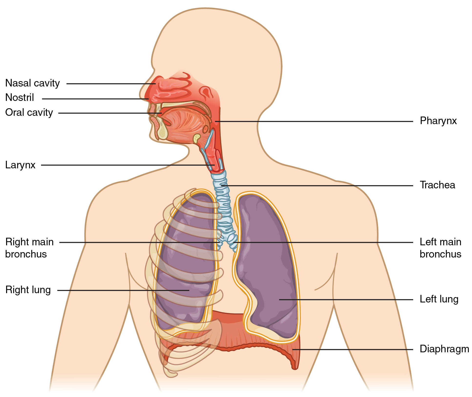
The major organs of the respiratory system function primarily to provide oxygen to body tissues for cellular respiration, remove the waste product carbon dioxide, and help to maintain acid-base balance. Portions of the respiratory system are also used for non-vital functions, such as sensing odors, speech production, and for straining, such as during childbirth or coughing (Figure 1).
Figure 1. Major Respiratory Structures. The major respiratory structures span the nasal cavity to the diaphragm.
Functionally, the respiratory system can be divided into a conducting zone and a respiratory zone. The conducting zone of the respiratory system includes the organs and structures not directly involved in gas exchange. The gas exchange occurs in the respiratory zone.
Conducting Zone
The major functions of the conducting zone are to provide a route for incoming and outgoing air, remove debris and pathogens from the incoming air, and warm and humidify the incoming air. Several structures within the conducting zone perform other functions as well. The epithelium of the nasal passages, for example, is essential to sensing odors, and the bronchial epithelium that lines the lungs can metabolize some airborne carcinogens.
The Nose and its Adjacent Structures
The major entrance and exit for the respiratory system is through the nose. When discussing the nose, it is helpful to divide it into two major sections: the external nose, and the nasal cavity or internal nose.
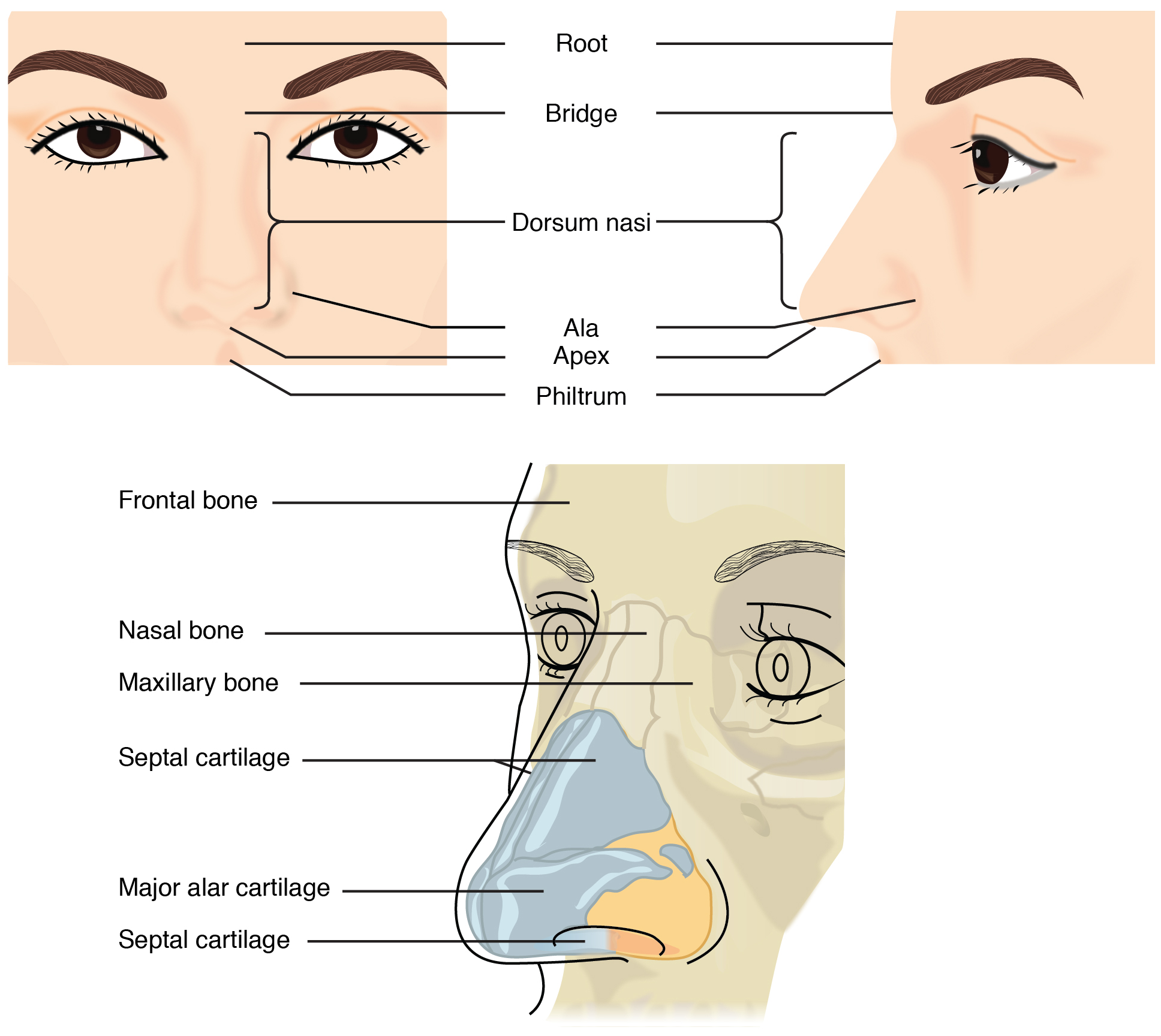
The external nose consists of the surface and skeletal structures that result in the outward appearance of the nose and contribute to its numerous functions (Figure 2). The root is the region of the nose located between the eyebrows. The bridge is the part of the nose that connects the root to the rest of the nose. The dorsum nasi is the length of the nose. The apex is the tip of the nose. On either side of the apex, the nostrils are formed by the alae (singular = ala). An ala is a cartilaginous structure that forms the lateral side of each naris (plural = nares), or nostril opening. The philtrum is the concave surface that connects the apex of the nose to the upper lip.
Figure 2. Nose. This illustration shows features of the external nose (top) and skeletal features of the nose (bottom).
Underneath the thin skin of the nose are its skeletal features (see Figure 2, lower illustration). While the root and bridge of the nose consist of bone, the protruding portion of the nose is composed of cartilage. As a result, when looking at a skull, the nose is missing. The nasal bone is one of a pair of bones that lies under the root and bridge of the nose. The nasal bone articulates superiorly with the frontal bone and laterally with the maxillary bones. Septal cartilage is flexible hyaline cartilage connected to the nasal bone, forming the dorsum nasi. The alar cartilage consists of the apex of the nose; it surrounds the naris.
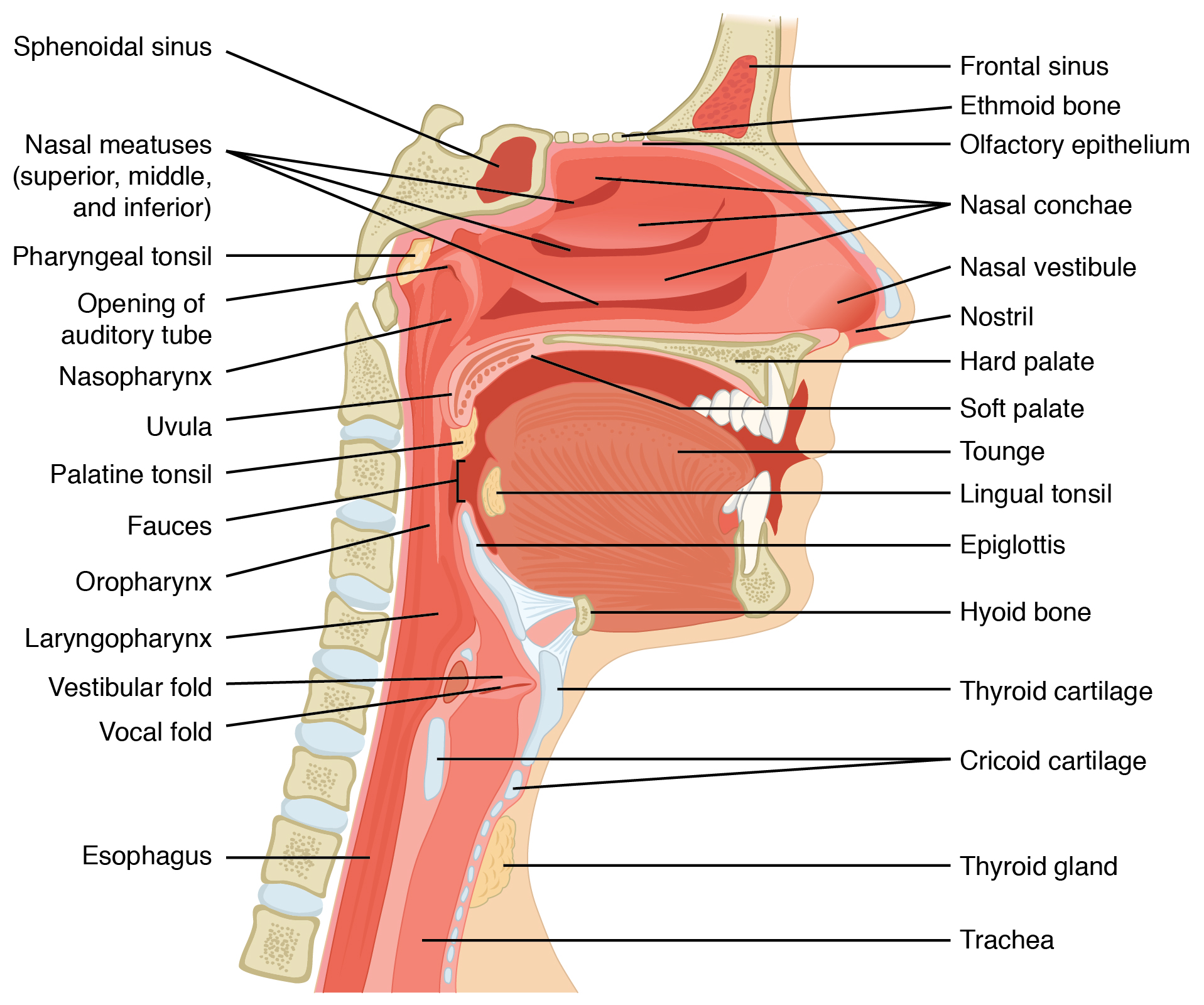
The nares open into the nasal cavity, which is separated into left and right sections by the nasal septum (Figure 3). The nasal septum is formed anteriorly by a portion of the septal cartilage (the flexible portion you can touch with your fingers) and posteriorly by the perpendicular plate of the ethmoid bone (a cranial bone located just posterior to the nasal bones) and the thin vomer bones (whose name refers to its plough shape). Each lateral wall of the nasal cavity has three bony projections, called the superior, middle, and inferior nasal conchae. The inferior conchae are separate bones, whereas the superior and middle conchae are portions of the ethmoid bone. Conchae serve to increase the surface area of the nasal cavity and to disrupt the flow of air as it enters the nose, causing air to bounce along the epithelium, where it is cleaned and warmed. The conchae and meatuses also conserve water and prevent dehydration of the nasal epithelium by trapping water during exhalation. The floor of the nasal cavity is composed of the palate. The hard palate at the anterior region of the nasal cavity is composed of bone. The soft palate at the posterior portion of the nasal cavity consists of muscle tissue. Air exits the nasal cavities via the internal nares and moves into the pharynx.
Figure 3. Upper Airway
Several bones that help form the walls of the nasal cavity have air-containing spaces called the paranasal sinuses, which serve to warm and humidify incoming air. Sinuses are lined with a mucosa. Each paranasal sinus is named for its associated bone: frontal sinus, maxillary sinus, sphenoidal sinus, and ethmoidal sinus. The sinuses produce mucus and lighten the weight of the skull.
The nares and anterior portion of the nasal cavities are lined with mucous membranes, containing sebaceous glands and hair follicles that serve to prevent the passage of large debris, such as dirt, through the nasal cavity. An olfactory epithelium used to detect odors is found deeper in the nasal cavity.
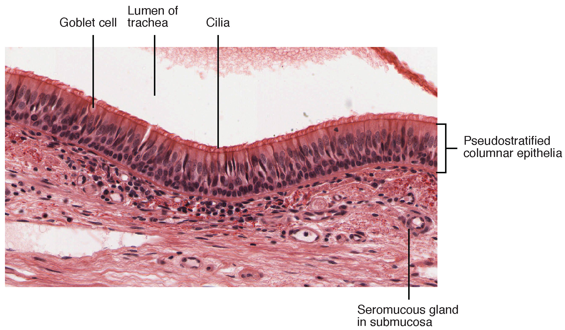
The conchae, meatuses, and paranasal sinuses are lined by respiratory epithelium composed of pseudostratified ciliated columnar epithelium (Figure 4). The epithelium contains goblet cells, one of the specialized, columnar epithelial cells that produce mucus to trap debris. The cilia of the respiratory epithelium help remove the mucus and debris from the nasal cavity with a constant beating motion, sweeping materials towards the throat to be swallowed. Interestingly, cold air slows the movement of the cilia, resulting in accumulation of mucus that may in turn lead to a runny nose during cold weather. This moist epithelium functions to warm and humidify incoming air. Capillaries located just beneath the nasal epithelium warm the air by convection. Serous and mucus-producing cells also secrete the lysozyme enzyme and proteins called defensins, which have antibacterial properties. Immune cells that patrol the connective tissue deep to the respiratory epithelium provide additional protection.
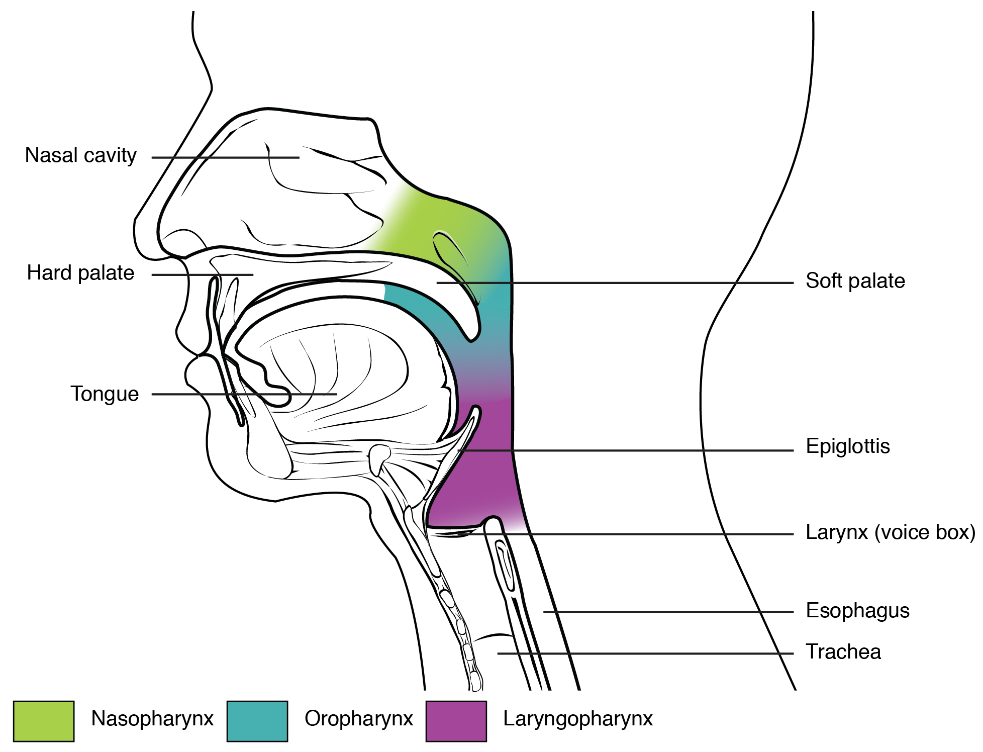
The pharynx is a tube formed by skeletal muscle and lined by mucous membrane that is continuous with that of the nasal cavities (see Figure 3). The pharynx is divided into three major regions: the nasopharynx, the oropharynx, and the laryngopharynx (Figure 5).
Figure 5. Divisions of the Pharynx. The pharynx is divided into three regions: the nasopharynx, the oropharynx, and the laryngopharynx.
The nasopharynx is flanked by the conchae of the nasal cavity, and it serves only as an airway. At the top of the nasopharynx are the pharyngeal tonsils. A pharyngeal tonsil, also called an adenoid, is an aggregate of lymphoid reticular tissue similar to a lymph node that lies at the superior portion of the nasopharynx. The function of the pharyngeal tonsil is not well understood, but it contains a rich supply of lymphocytes and is covered with ciliated epithelium that traps and destroys invading pathogens that enter during inhalation. The pharyngeal tonsils are large in children, but interestingly, tend to regress with age and may even disappear. The uvula is a small bulbous, teardrop-shaped structure located at the apex of the soft palate. Both the uvula and soft palate move like a pendulum during swallowing, swinging upward to close off the nasopharynx to prevent ingested materials from entering the nasal cavity. In addition, auditory (Eustachian) tubes that connect to each middle ear cavity open into the nasopharynx. This connection is why colds often lead to ear infections.
The oropharynx is a passageway for both air and food. The oropharynx is bordered superiorly by the nasopharynx and anteriorly by the oral cavity. The fauces is the opening at the connection between the oral cavity and the oropharynx. As the nasopharynx becomes the oropharynx, the epithelium changes from pseudostratified ciliated columnar epithelium to stratified squamous epithelium. The oropharynx contains two distinct sets of tonsils, the palatine and lingual tonsils. A palatine tonsil is one of a pair of structures located laterally in the oropharynx in the area of the fauces. The lingual tonsil is located at the base of the tongue. Similar to the pharyngeal tonsil, the palatine and lingual tonsils are composed of lymphoid tissue, and trap and destroy pathogens entering the body through the oral or nasal cavities.
The laryngopharynx is inferior to the oropharynx and posterior to the larynx. It continues the route for ingested material and air until its inferior end, where the digestive and respiratory systems diverge. The stratified squamous epithelium of the oropharynx is continuous with the laryngopharynx. Anteriorly, the laryngopharynx opens into the larynx, whereas posteriorly, it enters the esophagus.
Larynx
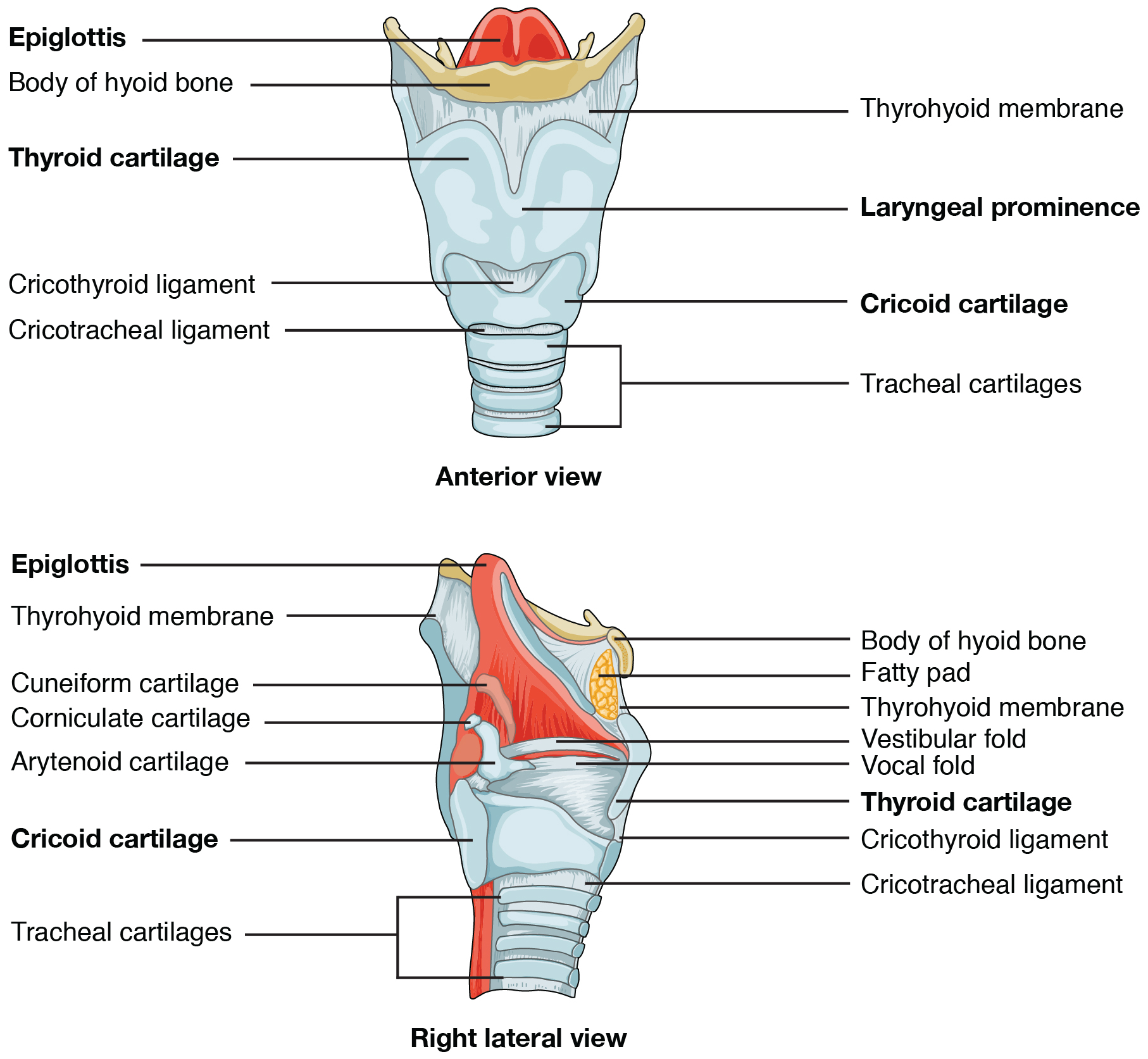
The larynx is a cartilaginous structure inferior to the laryngopharynx that connects the pharynx to the trachea and helps regulate the volume of air that enters and leaves the lungs (Figure 6). The structure of the larynx is formed by several pieces of cartilage. Three large cartilage pieces—the thyroid cartilage (anterior), epiglottis (superior), and cricoid cartilage (inferior)—form the major structure of the larynx. The thyroid cartilage is the largest piece of cartilage that makes up the larynx. The thyroid cartilage consists of the laryngeal prominence, or “Adam’s apple,” which tends to be more prominent in males. The thick cricoid cartilage forms a ring, with a wide posterior region and a thinner anterior region. Three smaller, paired cartilages—the arytenoids, corniculates, and cuneiforms—attach to the epiglottis and the vocal cords and muscle that help move the vocal cords to produce speech.
Figure 6. Larynx. The larynx extends from the laryngopharynx and the hyoid bone to the trachea.
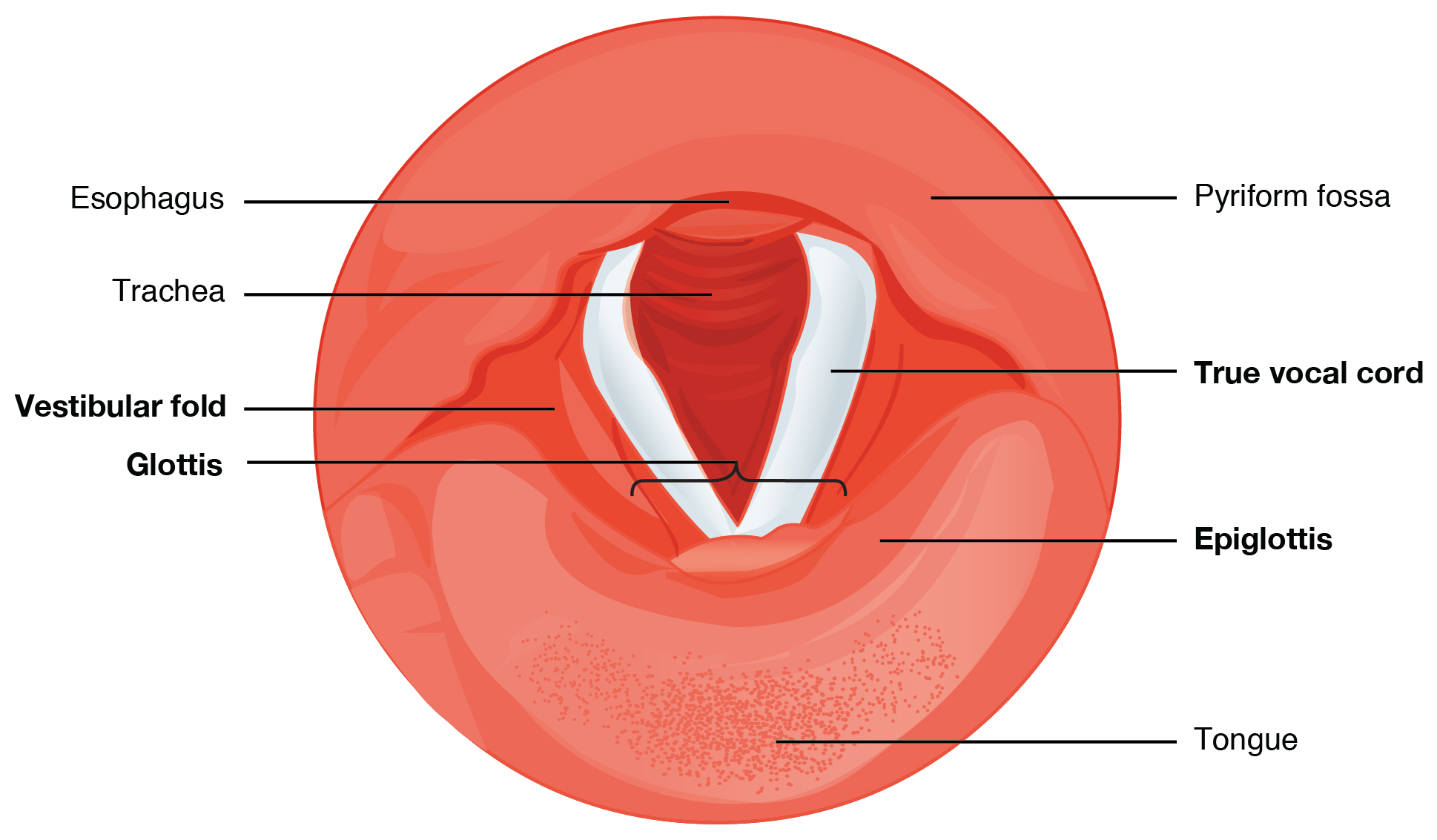
The epiglottis, attached to the thyroid cartilage, is a very flexible piece of elastic cartilage that covers the opening of the trachea (see Figure 3). When in the “closed” position, the unattached end of the epiglottis rests on the glottis. The glottis is composed of the vestibular folds, the true vocal cords, and the space between these folds (Figure 7). A vestibular fold, or false vocal cord, is one of a pair of folded sections of mucous membrane. A true vocal cord is one of the white, membranous folds attached by muscle to the thyroid and arytenoid cartilages of the larynx on their outer edges. The inner edges of the true vocal cords are free, allowing oscillation to produce sound. The size of the membranous folds of the true vocal cords differs between individuals, producing voices with different pitch ranges. Folds in males tend to be larger than those in females, which create a deeper voice. The act of swallowing causes the pharynx and larynx to lift upward, allowing the pharynx to expand and the epiglottis of the larynx to swing downward, closing the opening to the trachea. These movements produce a larger area for food to pass through, while preventing food and beverages from entering the trachea.
Figure 7. Vocal Cords. The true vocal cords and vestibular folds of the larynx are viewed inferiorly from the laryngopharynx.
Continuous with the laryngopharynx, the superior portion of the larynx is lined with stratified squamous epithelium, transitioning into pseudostratified ciliated columnar epithelium that contains goblet cells. Similar to the nasal cavity and nasopharynx, this specialized epithelium produces mucus to trap debris and pathogens as they enter the trachea. The cilia beat the mucus upward towards the laryngopharynx, where it can be swallowed down the esophagus.
Trachea
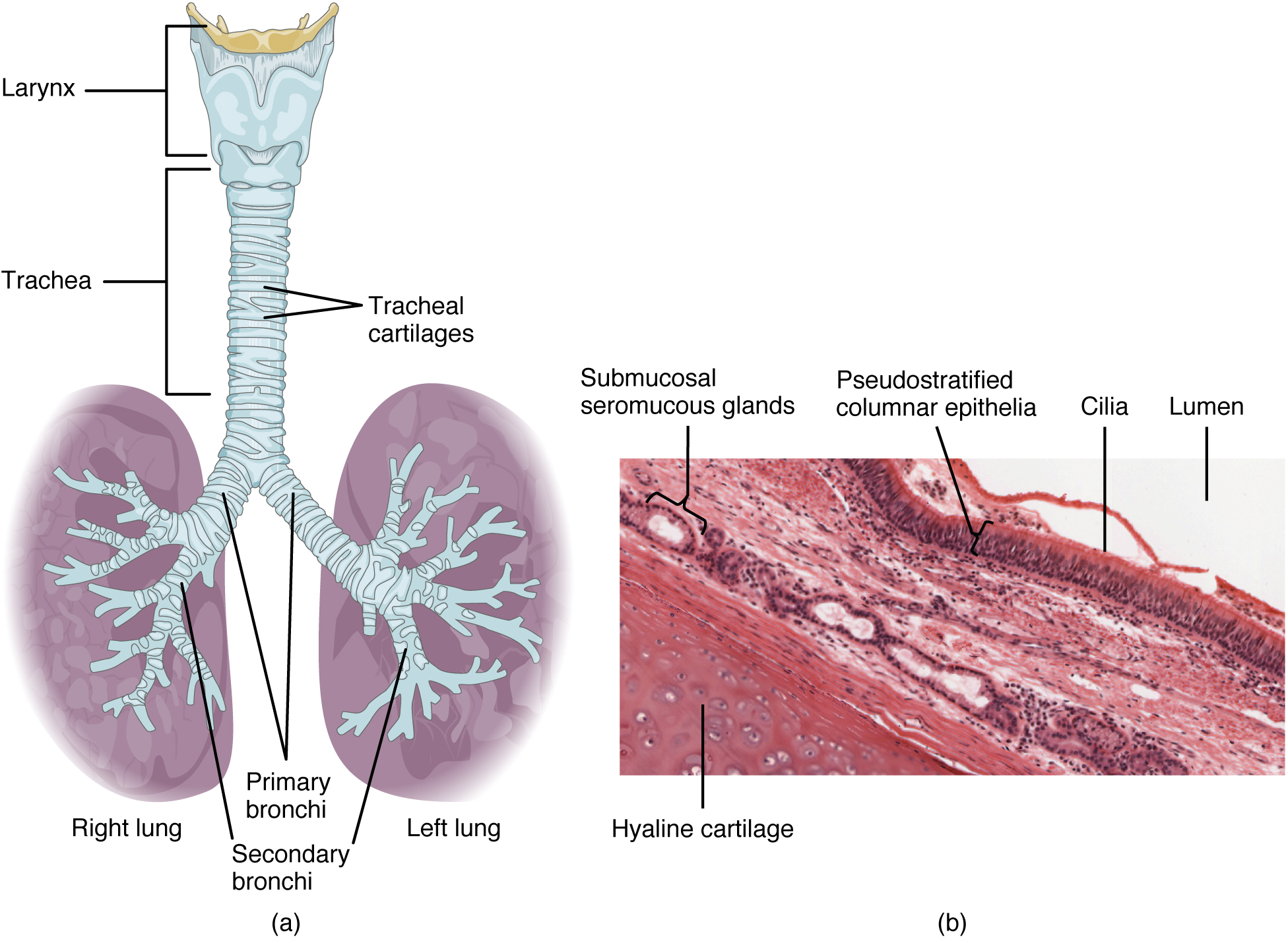
The trachea (windpipe) extends from the larynx toward the lungs (Figure 8a). The trachea is formed by 16 to 20 stacked, C-shaped pieces of hyaline cartilage that are connected by dense connective tissue. The trachealis muscle and elastic connective tissue together form the fibroelastic membrane, a flexible membrane that closes the posterior surface of the trachea, connecting the C-shaped cartilages. The fibroelastic membrane allows the trachea to stretch and expand slightly during inhalation and exhalation, whereas the rings of cartilage provide structural support and prevent the trachea from collapsing. In addition, the trachealis muscle can be contracted to force air through the trachea during exhalation. The trachea is lined with pseudostratified ciliated columnar epithelium, which is continuous with the larynx. The esophagus borders the trachea posteriorly.
The trachea branches into the right and left primary bronchi at the carina. These bronchi are also lined by pseudostratified ciliated columnar epithelium containing mucus-producing goblet cells (Figure 8b). The carina is a raised structure that contains specialized nervous tissue that induces violent coughing if a foreign body, such as food, is present. Rings of cartilage, similar to those of the trachea, support the structure of the bronchi and prevent their collapse. The primary bronchi enter the lungs at the hilum, a concave region where blood vessels, lymphatic vessels, and nerves also enter the lungs. The bronchi continue to branch into bronchial a tree. A bronchial tree (or respiratory tree) is the collective term used for these multiple-branched bronchi. The main function of the bronchi, like other conducting zone structures, is to provide a passageway for air to move into and out of each lung. In addition, the mucous membrane traps debris and pathogens.
A bronchiole branches from the tertiary bronchi. Bronchioles, which are about 1 mm in diameter, further branch until they become the tiny terminal bronchioles, which lead to the structures of gas exchange. There are more than 1000 terminal bronchioles in each lung. The muscular walls of the bronchioles do not contain cartilage like those of the bronchi. This muscular wall can change the size of the tubing to increase or decrease airflow through the tube.
Respiratory Zone
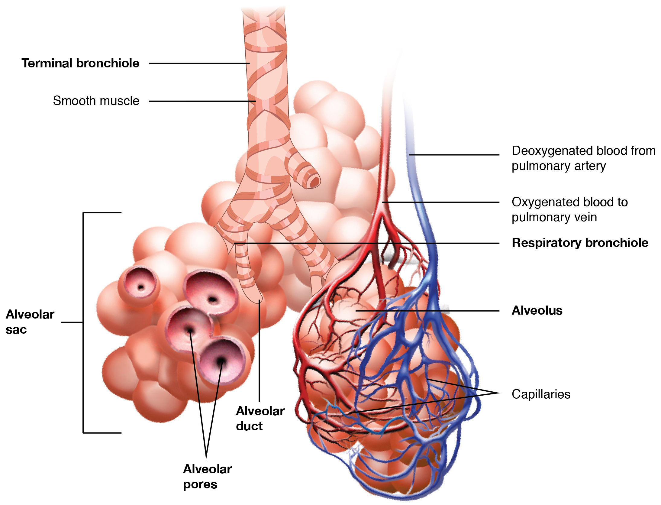
In contrast to the conducting zone, the respiratory zone includes structures that are directly involved in gas exchange. The respiratory zone begins where the terminal bronchioles join a respiratory bronchiole, the smallest type of bronchiole (Figure 9), which then leads to an alveolar duct, opening into a cluster of alveoli.
Figure 9. Respiratory Zone. Bronchioles lead to alveolar sacs in the respiratory zone, where gas exchange occurs.
Alveoli
An alveolar duct is a tube composed of smooth muscle and connective tissue, which opens into a cluster of alveoli. An alveolus is one of the many small, grape-like sacs that are attached to the alveolar ducts.
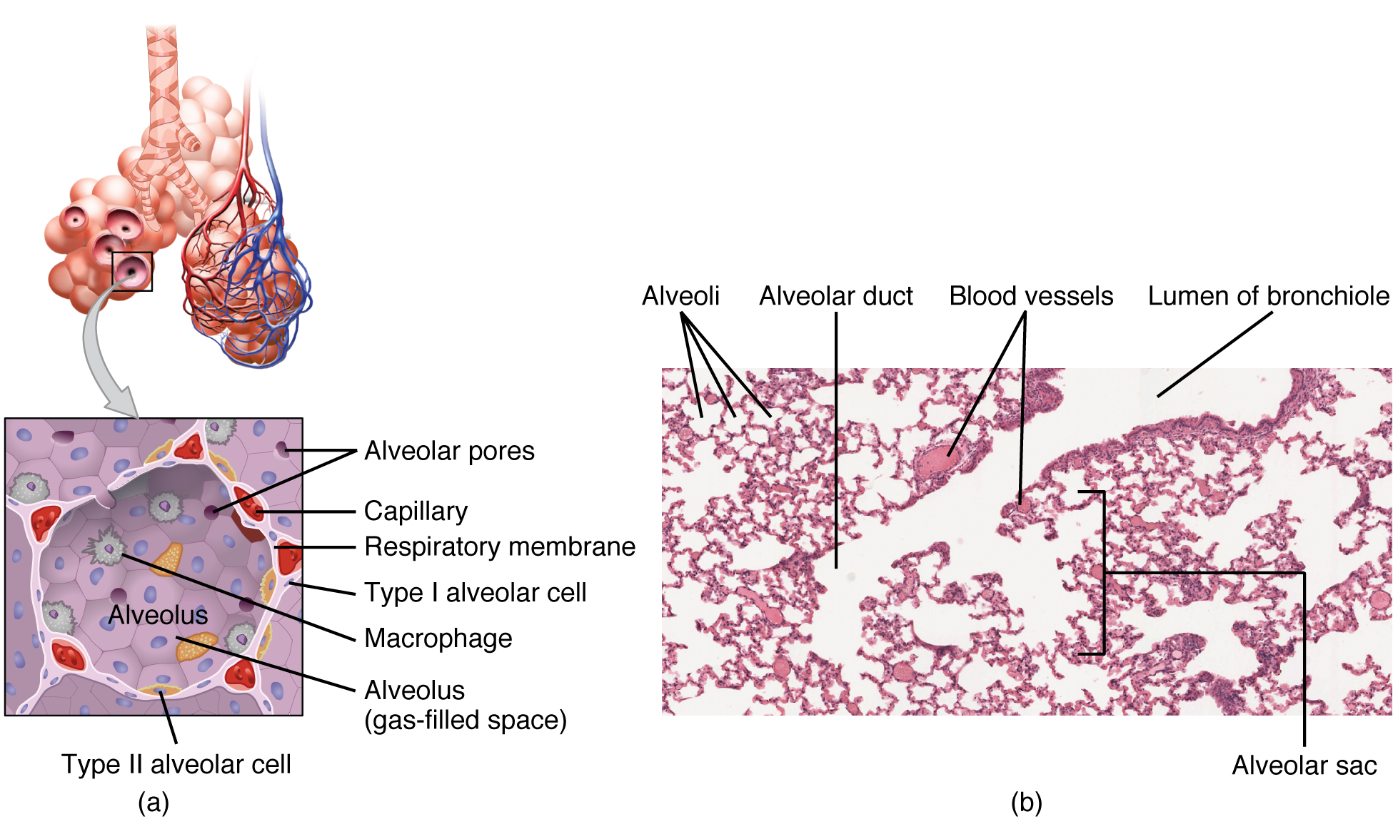
An alveolar sac is a cluster of many individual alveoli that are responsible for gas exchange. An alveolus is approximately 200 μm in diameter with elastic walls that allow the alveolus to stretch during air intake, which greatly increases the surface area available for gas exchange. Alveoli are connected to their neighbors by alveolar pores, which help maintain equal air pressure throughout the alveoli and lung (Figure 10).
The alveolar wall consists of three major cell types: type I alveolar cells, type II alveolar cells, and alveolar macrophages. A type I alveolar cell is a squamous epithelial cell of the alveoli, which constitute up to 97 percent of the alveolar surface area. These cells are about 25 nm thick and are highly permeable to gases. A type II alveolar cell is interspersed among the type I cells and secretes pulmonary surfactant, a substance composed of phospholipids and proteins that reduces the surface tension of the alveoli. Roaming around the alveolar wall is the alveolar macrophage, a phagocytic cell of the immune system that removes debris and pathogens that have reached the alveoli.
The simple squamous epithelium formed by type I alveolar cells is attached to a thin, elastic basement membrane. This epithelium is extremely thin and borders the endothelial membrane of capillaries. Taken together, the alveoli and capillary membranes form a respiratory membrane that is approximately 0.5 mm thick. The respiratory membrane allows gases to cross by simple diffusion, allowing oxygen to be picked up by the blood for transport and CO2to be released into the air of the alveoli.
Diseases of the…
Respiratory System: Asthma
Asthma is common condition that affects the lungs in both adults and children. Approximately 8.2 percent of adults (18.7 million) and 9.4 percent of children (7 million) in the United States suffer from asthma. In addition, asthma is the most frequent cause of hospitalization in children.
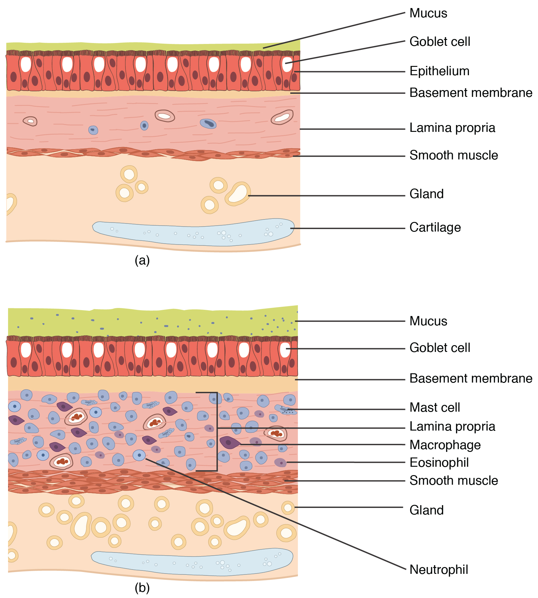
Asthma is a chronic disease characterized by inflammation and edema of the airway, and bronchospasms (that is, constriction of the bronchioles), which can inhibit air from entering the lungs. In addition, excessive mucus secretion can occur, which further contributes to airway occlusion (Figure 11). Cells of the immune system, such as eosinophils and mononuclear cells, may also be involved in infiltrating the walls of the bronchi and bronchioles.
Bronchospasms occur periodically and lead to an “asthma attack.” An attack may be triggered by environmental factors such as dust, pollen, pet hair, or dander, changes in the weather, mold, tobacco smoke, and respiratory infections, or by exercise and stress.
Figure 11. Normal and Bronchial Asthma Tissues. (a) Normal lung tissue does not have the characteristics of lung tissue during (b) an asthma attack, which include thickened mucosa, increased mucus-producing goblet cells, and eosinophil infiltrates.
Symptoms of an asthma attack involve coughing, shortness of breath, wheezing, and tightness of the chest. Symptoms of a severe asthma attack that requires immediate medical attention would include difficulty breathing that results in blue (cyanotic) lips or face, confusion, drowsiness, a rapid pulse, sweating, and severe anxiety. The severity of the condition, frequency of attacks, and identified triggers influence the type of medication that an individual may require. Longer-term treatments are used for those with more severe asthma. Short-term, fast-acting drugs that are used to treat an asthma attack are typically administered via an inhaler. For young children or individuals who have difficulty using an inhaler, asthma medications can be administered via a nebulizer.
In many cases, the underlying cause of the condition is unknown. However, recent research has demonstrated that certain viruses, such as human rhinovirus C (HRVC), and the bacteria Mycoplasma pneumoniae and Chlamydia pneumoniae that are contracted in infancy or early childhood, may contribute to the development of many cases of asthma.
Visit this site to learn more about what happens during an asthma attack.
Visit this site to learn more about what happens during an asthma attack. What are the three changes that occur inside the airways during an asthma attack?
Chapter Review
The respiratory system is responsible for obtaining oxygen and getting rid of carbon dioxide, and aiding in speech production and in sensing odors. From a functional perspective, the respiratory system can be divided into two major areas: the conducting zone and the respiratory zone. The conducting zone consists of all of the structures that provide passageways for air to travel into and out of the lungs: the nasal cavity, pharynx, trachea, bronchi, and most bronchioles. The nasal passages contain the conchae and meatuses that expand the surface area of the cavity, which helps to warm and humidify incoming air, while removing debris and pathogens. The pharynx is composed of three major sections: the nasopharynx, which is continuous with the nasal cavity; the oropharynx, which borders the nasopharynx and the oral cavity; and the laryngopharynx, which borders the oropharynx, trachea, and esophagus. The respiratory zone includes the structures of the lung that are directly involved in gas exchange: the terminal bronchioles and alveoli.
The lining of the conducting zone is composed mostly of pseudostratified ciliated columnar epithelium with goblet cells. The mucus traps pathogens and debris, whereas beating cilia move the mucus superiorly toward the throat, where it is swallowed. As the bronchioles become smaller and smaller, and nearer the alveoli, the epithelium thins and is simple squamous epithelium in the alveoli. The endothelium of the surrounding capillaries, together with the alveolar epithelium, forms the respiratory membrane. This is a blood-air barrier through which gas exchange occurs by simple diffusion.
Interactive Link Questions
Visit this site to learn more about what happens during an asthma attack. What are the three changes that occur inside the airways during an asthma attack?
Inflammation and the production of a thick mucus; constriction of the airway muscles, or bronchospasm; and an increased sensitivity to allergens.
Review Questions
1. Which of the following anatomical structures is not part of the conducting zone?
pharynx
nasal cavity
alveoli
bronchi
2. What is the function of the conchae in the nasal cavity?
increase surface area
exchange gases
maintain surface tension
maintain air pressure
3. The fauces connects which of the following structures to the oropharynx?
nasopharynx
laryngopharynx
nasal cavity
oral cavity
4. Which of the following are structural features of the trachea?
C-shaped cartilage
smooth muscle fibers
cilia
all of the above
5. Which of the following structures is not part of the bronchial tree?
alveoli
bronchi
terminal bronchioles
respiratory bronchioles
6. What is the role of alveolar macrophages?
to secrete pulmonary surfactant
to secrete antimicrobial proteins
to remove pathogens and debris
to facilitate gas exchange
Critical Thinking Questions
1. Describe the three regions of the pharynx and their functions.
2. If a person sustains an injury to the epiglottis, what would be the physiological result?
3. Compare and contrast the conducting and respiratory zones.
References
Bizzintino J, Lee WM, Laing IA, Vang F, Pappas T, Zhang G, Martin AC, Khoo SK, Cox DW, Geelhoed GC, et al. Association between human rhinovirus C and severity of acute asthma in children. Eur Respir J [Internet]. 2010 [cited 2013 Mar 22]; 37(5):1037–1042. Available from: http://erj.ersjournals.com/gca?submit=Go&gca=erj%3B37%2F5%2F1037&allch=
Kumar V, Ramzi S, Robbins SL. Robbins Basic Pathology. 7th ed. Philadelphia (PA): Elsevier Ltd; 2005.
(plural = alae) small, flaring structure of a nostril that forms the lateral side of the nares
alar cartilage
cartilage that supports the apex of the nose and helps shape the nares; it is connected to the septal cartilage and connective tissue of the alae
alveolar duct
small tube that leads from the terminal bronchiole to the respiratory bronchiole and is the point of attachment for alveoli
alveolar macrophage
immune system cell of the alveolus that removes debris and pathogens
alveolar pore
opening that allows airflow between neighboring alveoli
alveolar sac
cluster of alveoli
alveolus
small, grape-like sac that performs gas exchange in the lungs
apex
tip of the external nose
bronchial tree
collective name for the multiple branches of the bronchi and bronchioles of the respiratory system
bridge
portion of the external nose that lies in the area of the nasal bones
bronchiole
branch of bronchi that are 1 mm or less in diameter and terminate at alveolar sacs
bronchus
tube connected to the trachea that branches into many subsidiaries and provides a passageway for air to enter and leave the lungs
conducting zone
region of the respiratory system that includes the organs and structures that provide passageways for air and are not directly involved in gas exchange
cricoid cartilage
portion of the larynx composed of a ring of cartilage with a wide posterior region and a thinner anterior region; attached to the esophagus
dorsum nasi
intermediate portion of the external nose that connects the bridge to the apex and is supported by the nasal bone
epiglottis
leaf-shaped piece of elastic cartilage that is a portion of the larynx that swings to close the trachea during swallowing
external nose
region of the nose that is easily visible to others
fauces
portion of the posterior oral cavity that connects the oral cavity to the oropharynx
fibroelastic membrane
specialized membrane that connects the ends of the C-shape cartilage in the trachea; contains smooth muscle fibers
glottis
opening between the vocal folds through which air passes when producing speech
laryngeal prominence
region where the two lamina of the thyroid cartilage join, forming a protrusion known as “Adam’s apple”
laryngopharynx
portion of the pharynx bordered by the oropharynx superiorly and esophagus and trachea inferiorly; serves as a route for both air and food
larynx
cartilaginous structure that produces the voice, prevents food and beverages from entering the trachea, and regulates the volume of air that enters and leaves the lungs
lingual tonsil
lymphoid tissue located at the base of the tongue
meatus
one of three recesses (superior, middle, and inferior) in the nasal cavity attached to the conchae that increase the surface area of the nasal cavity
naris
(plural = nares) opening of the nostrils
nasal bone
bone of the skull that lies under the root and bridge of the nose and is connected to the frontal and maxillary bones
nasal septum
wall composed of bone and cartilage that separates the left and right nasal cavities
nasopharynx
portion of the pharynx flanked by the conchae and oropharynx that serves as an airway
oropharynx
portion of the pharynx flanked by the nasopharynx, oral cavity, and laryngopharynx that is a passageway for both air and food
palatine tonsil
one of the paired structures composed of lymphoid tissue located anterior to the uvula at the roof of isthmus of the fauces
paranasal sinus
one of the cavities within the skull that is connected to the conchae that serve to warm and humidify incoming air, produce mucus, and lighten the weight of the skull; consists of frontal, maxillary, sphenoidal, and ethmoidal sinuses
pharyngeal tonsil
structure composed of lymphoid tissue located in the nasopharynx
pharynx
region of the conducting zone that forms a tube of skeletal muscle lined with respiratory epithelium; located between the nasal conchae and the esophagus and trachea
philtrum
concave surface of the face that connects the apex of the nose to the top lip
pulmonary surfactant
substance composed of phospholipids and proteins that reduces the surface tension of the alveoli; made by type II alveolar cells
respiratory bronchiole
specific type of bronchiole that leads to alveolar sacs
respiratory epithelium
ciliated lining of much of the conducting zone that is specialized to remove debris and pathogens, and produce mucus
respiratory membrane
alveolar and capillary wall together, which form an air-blood barrier that facilitates the simple diffusion of gases
respiratory zone
includes structures of the respiratory system that are directly involved in gas exchange
root
region of the external nose between the eyebrows
thyroid cartilage
largest piece of cartilage that makes up the larynx and consists of two lamina
trachea
tube composed of cartilaginous rings and supporting tissue that connects the lung bronchi and the larynx; provides a route for air to enter and exit the lung
trachealis muscle
smooth muscle located in the fibroelastic membrane of the trachea
true vocal cord
one of the pair of folded, white membranes that have a free inner edge that oscillates as air passes through to produce sound
type I alveolar cell
squamous epithelial cells that are the major cell type in the alveolar wall; highly permeable to gases
type II alveolar cell
cuboidal epithelial cells that are the minor cell type in the alveolar wall; secrete pulmonary surfactant
vestibular fold
part of the folded region of the glottis composed of mucous membrane; supports the epiglottis during swallowing
Solutions
Answers for Review Questions
C
A
D
A
C
C
Answers for Critical Thinking Questions
The pharynx has three major regions. The first region is the nasopharynx, which is connected to the posterior nasal cavity and functions as an airway. The second region is the oropharynx, which is continuous with the nasopharynx and is connected to the oral cavity at the fauces. The laryngopharynx is connected to the oropharynx and the esophagus and trachea. Both the oropharynx and laryngopharynx are passageways for air and food and drink.
The epiglottis is a region of the larynx that is important during the swallowing of food or drink. As a person swallows, the pharynx moves upward and the epiglottis closes over the trachea, preventing food or drink from entering the trachea. If a person’s epiglottis were injured, this mechanism would be impaired. As a result, the person may have problems with food or drink entering the trachea, and possibly, the lungs. Over time, this may cause infections such as pneumonia to set in.
The conducting zone of the respiratory system includes the organs and structures that are not directly involved in gas exchange, but perform other duties such as providing a passageway for air, trapping and removing debris and pathogens, and warming and humidifying incoming air. Such structures include the nasal cavity, pharynx, larynx, trachea, and most of the bronchial tree. The respiratory zone includes all the organs and structures that are directly involved in gas exchange, including the respiratory bronchioles, alveolar ducts, and alveoli.
LICENSE
he upper respiratory system includes the mouth, nose, nasal cavity, sinuses, and the pharynx. The lower respiratory system begins with the larynx, or voice box, and includes the trachea, or wind pipe, bronchi, bronchioles, and alveoli within the lungs
The airway, or respiratory tract, describes the organs of the respiratory tract that allow air flow during ventilation. They reach from the nares and buccal opening to the blind end of the alveolar sacs. They are subdivided into different regions with various organs and tissues to perform specific functions. The airway can be subdivided into the upper and lower airway, each of which has numerous subdivisions as follows.
Upper Airway
The pharynx is the mucous membrane-lined portion of the airway between the base of the skull and the esophagus and is subdivided as follows:
Nasopharynx, also known as the rhino-pharynx, post-nasal space, is the muscular tube from the nares, including the posterior nasal cavity, divide from the oropharynx by the palate and lining the skull base superiorly
The oro-pharynx connects the naso and hypopharynx. It is the region between the palate and the hyoid bone, anteriorly divided from the oral cavity by the tonsillar arch
The hypopharynx connects the oropharynx to the esophagus and the larynx, the region of pharynx below the hyoid bone.
The larynx is the portion of the airway between the pharynx and the trachea, contains the organs for production of speech. Formed of a cartilaginous skeleton of nine cartilages, it includes the important organs of the epiglottis and the vocal folds (vocal chords) which are the opening to the glottis.
Lower Airway
The trachea is a ciliated pseudostratified columnar epithelium-lined tubular structure supported by C-shaped rings of hyaline cartilage. The flat open surface of these C rings opposes the esophagus to allow its expansion during swallowing. The trachea bifurcates and therefore terminates, superior to the heart at the level of the sternal angle.
The bronchi, the main bifurcation of the trachea, are similar in structure but have complete circular cartilage rings.
Main bronchi: There are two supplying ventilation to each lung. The right main bronchus has a larger diameter and is aligned more vertically than the left
Lobar bronchi: Two on the left and three on the right supply each of the main lobes of the lung
Segmental bronchi supply individual bronchopulmonary segments of the lungs.
Bronchioles lack supporting cartilage skeletons and have a diameter of around 1 mm. They are initially ciliated and graduate to the simple columnar epithelium and their lining cells no longer contain mucous producing cells.
Conducting bronchioles conduct airflow but do not contain any mucous glands or seromucous glands
Terminal bronchioles are the last division of the airway without respiratory surfaces
Respiratory bronchioles contain occasional alveoli and have surface surfactant-producing They each give rise to between two and 11 alveolar ducts.
Alveolar is the final portion of the airway and is lined with a single-cell layer of pneumocytes and in proximity to capillaries. They contain surfactant producing type II pneumocytes and Clara cells.
Alveolar ducts are tubular portions with respiratory surfaces from which the alveolar sacs bud.
Alveolar sacs are the blind-ended spaces from which the alveoli clusters are formed and to where they connect. These are connected by pores which allow air pressure to equalize between them. Together, with the capillaries, they form the air-blood barrier.
Airways allow air flow in ventilation from the external environment to the respiratory surfaces where gas exchange for respiratory processes can occur.
To allow this and to maintain homeostasis and adequate protection from the external environment they must also perform other barrier functions.
Moisture barrier is the mucous lining of the airway that provides a barrier to prevent loss of excessive moisture during ventilation by increasing the humidity of the air in the upper airway
Temperature barrier is relative to body temperature as the external environment is nearly always colder, and the increased vasculature and structures such as nasal turbinates warm air as it enters the airways
A barrier to infection as the airways are lined with a rich lymphatic system including mucosa-associated lymphoid tissue (MALT) that prevents early access to any invading pathogens. Macrophages also patrol the respiratory surfaces providing an important component of the “air-blood barrier."
The upper airways develop from the pharyngeal arches as part of the embryological development of the head and neck structures.
At around four weeks, the larynx and lower airways develop from the longitudinal laryngotracheal groove which forms a medial, groove-like structure becoming a tubular, blind-ended structure called the laryngotracheal diverticulum. This eventually separates from the developing foregut by the formation of the tracheo-oesophageal folds.
The laryngeal cartilages and musculature develop from the four and six pharyngeal arches, and the glottic opening forms connecting this region to the trachea.
The trachea forms from the extension of the laryngotracheal diverticulum, and it is lined with endodermal tissue which forms the specialized respiratory linings and mesodermal structures which form the cartilage and smooth muscle walls.
As development continues, the laryngotracheal diverticulum continues to branch and bud and forms the bronchi and branching bronchioles.
From 16 weeks onward, respiratory surfaces begin to form, and the maturation of the lungs develops with alveolar sacs forming and pneumocyte development forming the respiratory membrane.
The formation of the alveoli and the respiratory membrane is not complete until after birth, and alveolar formation continues until the age of eight.
The upper airways receive blood supply from various branches of the external carotid artery and drain into the internal jugular. The naso and oro-pharynx also receive blood supply from the facial artery branch of the external carotid via the tonsillar artery. The venous drainage of these structures is via the pharyngeal plexus into the internal jugular vein. The lymphatic drainage is through various lymphatic plexuses of the neck surrounding the internal jugular vessels.
The lower airways receive blood flow from two sources: the pulmonary circulation and the bronchial circulation.
The pulmonary circulation provides blood from the heart for oxygenation through the right and left pulmonary arteries which follow a branching structure similar to that of the airways themselves. This blood returns as oxygenated blood through the pulmonary veins which follow an independently branching structure to return to the right ventricle.
Bronchial circulation provides oxygenated blood to the airway structures themselves. These arteries arise independently from the systemic circulation. The two left bronchial arteries emerge from the thoracic aorta; whereas, the right bronchial artery arises either from one of the superior posterior intercostal arteries or a common trunk with the left superior bronchial artery. These provide nutrition and oxygen to tissues as far as the end of the conducting airways where they anastomose with the pulmonary circulation.
The bronchial veins are only present near the lung hilum which drain blood from the trachea, and bronchi drain into the azygos vein on the right and either the accessory hemiazygos veins or the intercostal vessels on the left. Pulmonary veins drain the more distal circulation where a small amount of de-oxygenated blood makes a minimal impact on the saturation of the returning blood.
Lymphatic drainage of the lower airways is through the deep lymphatic plexuses of the pulmonary lymphatic plexuses. These drain to the superior and inferior tracheobronchial lymph nodes bilaterally and then to the right and left ducts connecting to the venous angles, usually directly but on the left, this may converge with the thoracic duct first.
Paratracheal nodes drain lymph from the trachea directly into the right and left lymphatic ducts.
Innervation of the pharynx is via cranial nerves VII, IX, X, and XII. The larynx is supplied by the vagus (cranial nerve X) by the superior laryngeal branch directly and the clinically important recurrent laryngeal branch.
The lower airways receive parasympathetic fibers from the vagus, some of which are afferent sensory nerves that transmit cough sensations from specialized J receptors in the mucosa as well as stretch receptors from the bronchial muscles and inter-alveolar connective tissues. The efferent fibers of the vagus cause broncho-constriction and secretion from the glandular tissues in the airways. The efferent sympathetic fibers cause bronchodilation by inhibiting the activity of the smooth muscles of the airways.
The muscles of the pharynx and larynx provide the structure of the upper airways and form from striated muscles under visceral and somatic control. They relate to the action of swallowing.
The lower airways have a layer of smooth muscle within their walls. It is present along all of the conducting airways and allows for visceral control of bronchoconstriction
The most common anatomic variation is an abnormal tracheo-oesophageal fistula. This variation occurs most commonly in males and often is associated with oesophageal atresia. It occurs with the incomplete fusion of the tracheo-oesophageal folds which would divide the developing foregut into respiratory and oesophageal portions.
The anatomy of the airway is important in all trauma and emergency surgical scenarios. As in any emergency assessment, a practitioner should know it is most important to consider and evaluate the patent airway.
The upper airways can be controlled using airway devices and bypassed using endotracheal intubation. If this is not possible, emergent surgical access to the airway is imperative and is performed through an emergency cricothyroidotomy.
Airway assessment is relevant to many common surgeries:
Tonsils causing any airway compromise indicate surgical removal.
Any neck trauma external to the airway can cause an external compression which can compromise the airway. This compromise is of particular importance in trauma and operations on surrounding structures such as thyroidectomy.
The importance of the upper airway assessment is paramount in both emergency and anesthetic scenarios.
The upper airway assessment can be performed and enhanced by the following assessment tools:
The Malampati score which describes the visible airway
The "3, 3, 2" rule in which three estimated measurements of the interincisor distance
The hyoid-mental distance, hyoid-thyroid cartilage distance is measured, and if these are shortened, it implies a difficult airway.
The cricoid cartilage is important both as a clinical landmark and also as the only complete cartilage ring within the upper airway used during cricoid pressure maneuvers.
The narrowest portion of the upper airway is the cricoid cartilage in children; therefore, cricothyroidotomy is not recommended in children younger than the age of eight. As children grow and mature, the glottic opening becomes the most narrow point in the airway, and therefore, the most likely point of obstruction and allow bypass by the insertion of a cricothyroidotomy airway.
The trachea is the most anterior structure of the neck except for where the thyroid covers it. This means that it can be accessed to provide an airway in both emergency situation (cricothyroidotomy) and elective procedures (tracheotomy).
The trachea should align with the sternal notch. If this alignment deviates, it can indicate a lung or mediastinal pathology.
The right, main bronchus is shorter, wider, and vertically aligned, and this means it is the most common site for aspiration, both in a foreign body aspiration and during the occurrence of an aspiration pneumonitis causing right lower lobe consolidation.
In clinical assessment of the lower airways, through auscultation and by the presence of "wheezing" as turbulent airflow generates a musical noise, airway narrowing through edema or bronchoconstriction can be detected.
My husband was diagnosed with early onset Parkinson's disease at 57.his symptoms were shuffling of feet,slurred speech, low volume speech, degradation of hand writing, horrible driving skills, right arm held at 45 degree angle, things were tough for me, but now he finally free from the disease with the help of total cure ultimate health home, he now walks properly and all symptoms has reversed, he had trouble with balance especially at night, getting into the shower and exiting it is difficult,getting into bed is also another thing he finds impossible.we had to find a better solution for his condition which has really helped him a lot,the biggest helped we had was ultimatehealthhome they walked us through the proper steps,am highly recommended this ultimatehealthhome@gmail.com to anyone who needs help.


My husband was diagnosed with early onset Parkinson's disease at 57.his symptoms were shuffling of feet,slurred speech, low volume speech, degradation of hand writing, horrible driving skills, right arm held at 45 degree angle, things were tough for me, but now he finally free from the disease with the help of total cure ultimate health home, he now walks properly and all symptoms has reversed, he had trouble with balance especially at night, getting into the shower and exiting it is difficult,getting into bed is also another thing he finds impossible.we had to find a better solution for his condition which has really helped him a lot,the biggest helped we had was ultimatehealthhome they walked us through the proper steps,am highly recommended this ultimatehealthhome@gmail.com to anyone who needs help.
ReplyDelete